3D printing has been used to produce rocket parts, medical implants and even to make art, and now 3D-printable models of anatomical specimens can be added to the list. A cane toad skeleton and part of a spiny dogfish skull are the first examples made available for students to download, with a view to making hands-on study more accessible than using biological specimens alone.
The pieces were created from original specimens using a 3D scanning and printing method developed by scientists at Massey University in New Zealand. The process uses consumer-level technology and commercial 3D printers, demonstrating how high-quality replicas can be easy and inexpensive substitutes where biological samples might be difficult to obtain.
Dr Daniel Thomas, lead author of a paper outlining the process published in the Journal of Anatomy, is excited by the prospect. "Imagine a classroom in Silverdale being able to print a moa skeleton or a university class in America being able to examine a kakapo beak that was scanned here in New Zealand," he said.
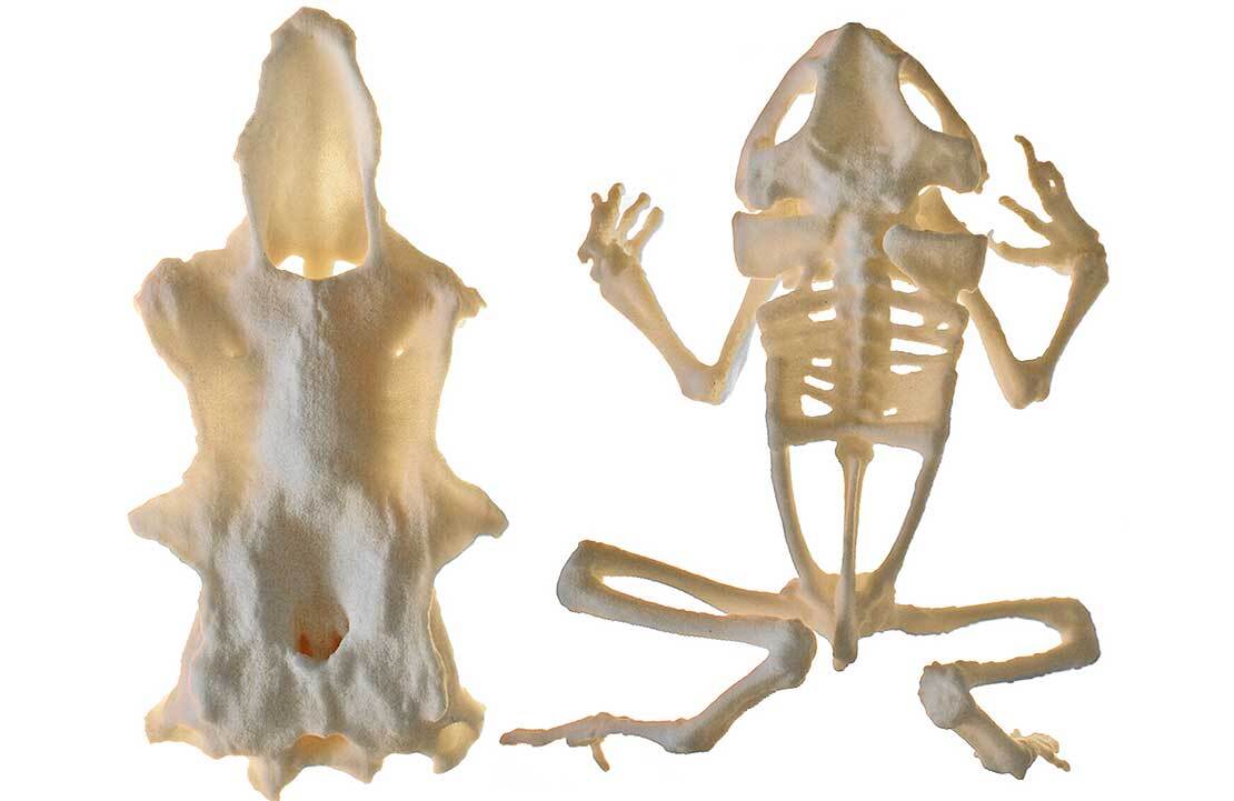
3D-printed copies aren't a total replacement for original specimens, which will display natural biological variation, but the level of detail on the models is impressive, with some dried muscle tissue reportedly visible on the vertebrae of the frog skeleton model.
Both the cane toad skeleton and the spiny dogfish skull models are available to download for free.
Source: Massey University