X-ray
-
 A supermassive monster lurks at the center of our galaxy, and astronomers have now discovered that it’s spinning so fast it’s warping the very fabric of spacetime into a football shape.
 -
 Astronomers have mapped out half the universe in X-ray light, using a space telescope called eROSITA. The new map, which contains almost a million X-ray sources, is the basis of dozens of new scientific papers, with many more to come.
 -
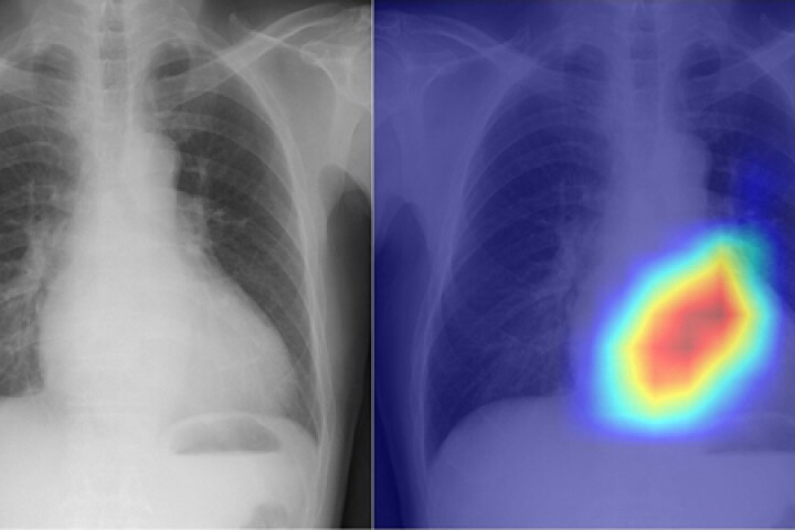
 Researchers developed an AI algorithm that automatically analyzes chest X-rays to rapidly detect COVID-19 infection with more than 98% accuracy, distinguishing between normal X-rays and X-rays from people with viral pneumonia.
 -
 Astronomers have discovered evidence of a theorized type of black hole lurking in the distant universe. Known as an “Outsize Black Hole,” this object could help explain some fundamental cosmic mysteries, including how supermassive monsters form.
 -
 As spooky season wraps, NASA has a final Halloween treat. Using two X-ray telescopes, astronomers have visualized the ‘bones’ in one of space's most haunting phenomena, the pulsar wind nebula MSH 15-52, also known as the ‘cosmic hand' or 'hand of God'.
 -
 The SLAC National Accelerator Laboratory has switched on the world's most powerful X-ray laser. The Linac Coherent Light Source II X-ray Free-Electron Laser (XFEL) flashes a million times a minute and is 10,000 times brighter than its predecessor.
 -
 It was second time lucky for Japan as it successfully launched its first lunar lander on September 6 at 23:42 GMT atop a Mitsubishi Heavy Industries H-IIA rocket from the Tanegashima Space Center 25 miles (40 km) south of Kyushu.
 -
 Using a deep-learning AI model, researchers have turned the humble chest X-ray into a more powerful tool for diagnosing heart problems. They say their novel approach could be used as a quick and accurate way of assessing heart function.
 -
 At the heart of our galaxy lurks a supermassive monster, currently slumbering. But new observations reveal an X-ray “echo” from a time when the Milky Way’s central black hole awoke just 200 years ago, shining a million times brighter than today.
 -
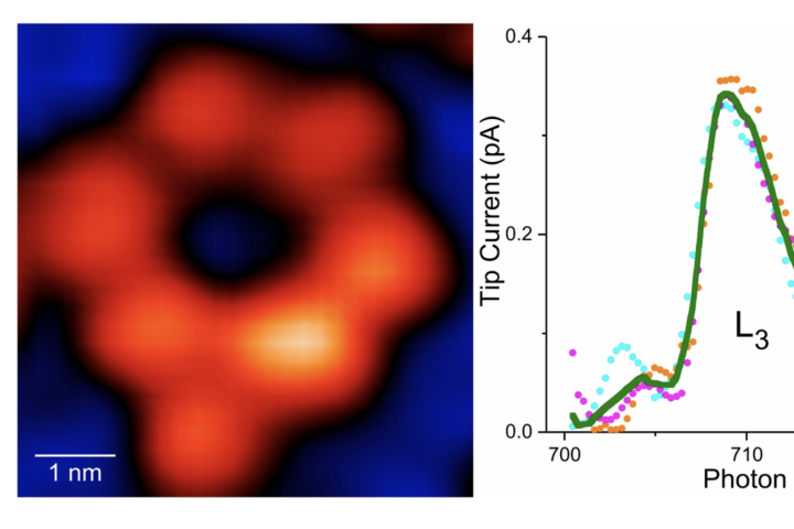 A team of researchers led by Ohio University Professor of Physics Saw Wai Hla has captured the first X-ray "image" of a single atom, allowing scientists to study materials and their chemical states with greater resolution than ever before.
 -
 The humble X-ray may have received a long-overdue upgrade thanks to the development of a highly sensitive, printable X-ray detector that can operate over a wide range of energy levels, with potential in a wide range of real-world applications.
 -
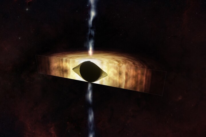
 Astronomers have spotted an incredibly bright flash of light beaming halfway across the universe. The strange light was estimated to throw off more light than one quadrillion Suns, and in an ironic twist came from one of the darkest objects possible.
Load More